
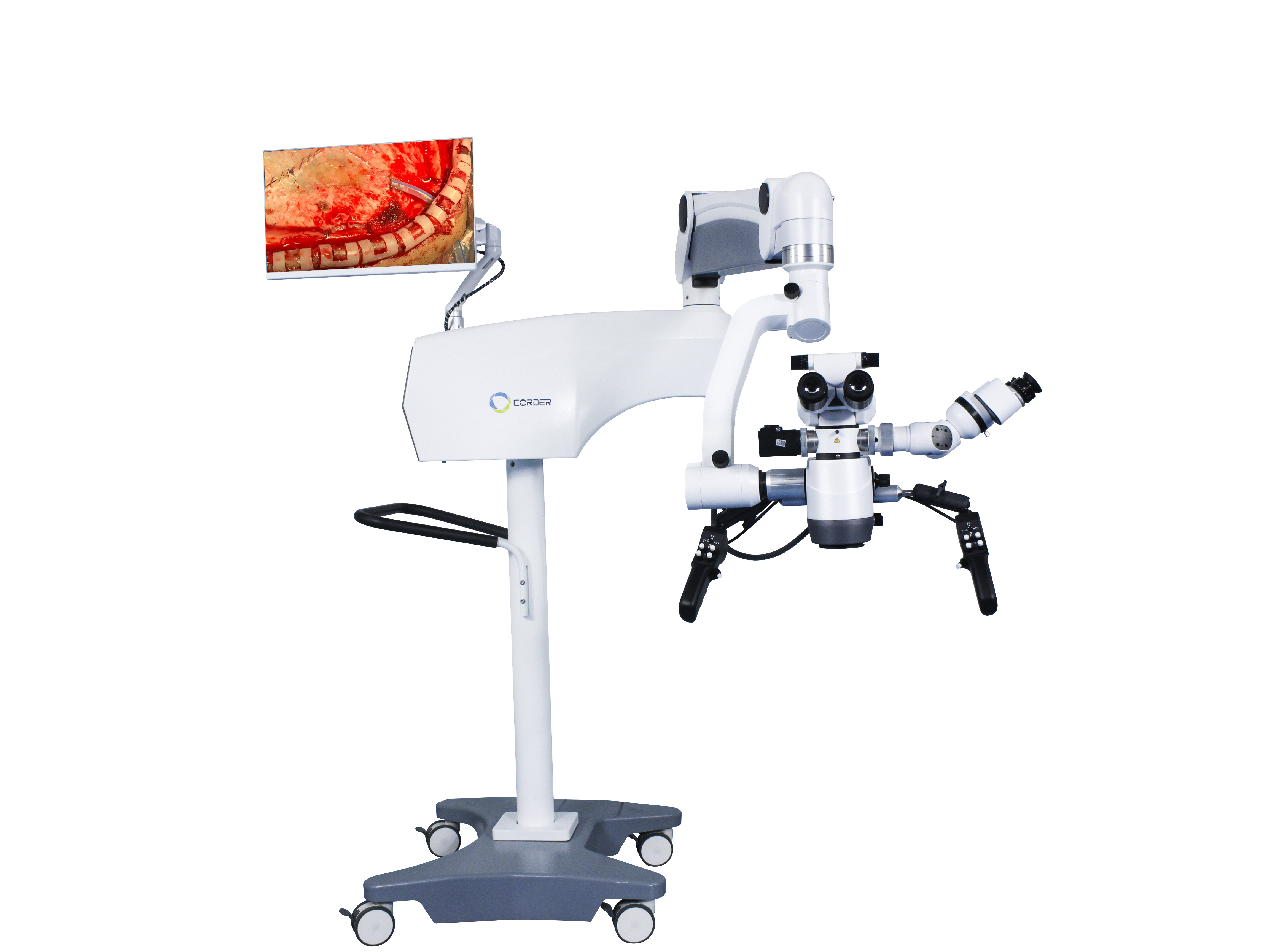
Neyroxirurgiyada jarrohlik mikroskoplarining qo'llanilish tarixi va roli
Neyroxirurgiya tarixida, qo'llanilishijarrohlik mikroskoplariyalang'och ko'z ostida jarrohlik amaliyotini o'tkazishning an'anaviy neyroxirurgik davridan zamonaviy neyroxirurgik davrga o'tayotgan inqilobiy ramzdir.mikroskopKim va qachon qildioperatsion mikroskoplarneyroxirurgiyada qo'llanila boshlandi? Qanday rol o'ynadijarrohlik mikroskopineyroxirurgiyaning rivojlanishida qanday rol o'ynagan? Fan va texnologiyaning rivojlanishi bilan,Operatsion mikroskopyanada ilg'or uskunalar bilan almashtirilishi mumkinmi? Bu har bir neyroxirurg bilishi va neyroxirurgiya sohasidagi eng so'nggi texnologiyalar va vositalarni qo'llashi, neyroxirurgiya jarrohlik ko'nikmalarini oshirishga yordam berishi kerak bo'lgan savol.
1, Tibbiyot sohasida mikroskopiya qo'llanilishi tarixi
Fizikada ko'zoynak linzalari - bu kattalashtirish effektiga ega bo'lgan bitta tuzilishga ega bo'lgan qavariq linzalar bo'lib, ularning kattalashtirishi cheklangan bo'lib, u kattalashtiruvchi ko'zoynaklar deb ataladi. 1590-yilda ikki gollandiyalik ingichka silindrsimon bochka ichiga ikkita qavariq linza plastinkasini o'rnatdilar va shu tariqa dunyodagi birinchi kompozit strukturali kattalashtirish moslamasini ixtiro qildilar:mikroskopKeyinchalik, mikroskopning tuzilishi doimiy ravishda takomillashtirildi va kattalashtirish doimiy ravishda oshdi. O'sha paytda olimlar asosan bundan foydalanishgankompozit mikroskophayvonlar va o'simliklarning mayda tuzilmalarini, masalan, hujayralar tuzilishini kuzatish uchun. 19-asrning o'rtalaridan oxirigacha tibbiyot sohasida kattalashtiruvchi ko'zoynaklar va mikroskoplar asta-sekin qo'llanila boshlandi. Dastlab, jarrohlar jarrohlik amaliyoti uchun burun ko'prigiga joylashtirilishi mumkin bo'lgan bitta linzali tuzilishga ega ko'zoynak uslubidagi kattalashtiruvchi ko'zoynaklardan foydalanishgan. 1876-yilda nemis shifokori Saemisch murakkab ko'zoynak kattalashtiruvchi oynasidan foydalanib (jarrohlik turi noma'lum) dunyodagi birinchi "mikroskopik" operatsiyani amalga oshirdi. 1893-yilda nemis Zeiss kompaniyasi ixtiro qildi...durbin mikroskopi, asosan tibbiy laboratoriyalarda eksperimental kuzatish uchun, shuningdek, oftalmologiya sohasida shox parda va oldingi kameraning shikastlanishlarini kuzatish uchun ishlatiladi. 1921-yilda hayvonlarning ichki qulog'i anatomiyasi bo'yicha laboratoriya tadqiqotlariga asoslanib, shved otolaringologi Nylen fiksatsiyalangan usuldan foydalangan.monokulyar jarrohlik mikroskopiOdamlarda surunkali otit media operatsiyasini o'tkazish uchun o'zi tomonidan ishlab chiqilgan va ishlab chiqarilgan, bu haqiqiy mikrojarrohlik edi. Bir yil o'tgach, Nylenning yuqori lavozimli shifokori Xlolmgren ... ni taqdim etdi.binokulyar jarrohlik mikroskopiZeiss tomonidan operatsiya xonasida ishlab chiqarilgan.
ErtaOperatsion mikroskoplarmexanik barqarorlikning pastligi, harakatlana olmaslik, turli o'qlarning yoritilishi va obyektiv linzaning qizishi, jarrohlik kattalashtirish maydonining torligi va boshqalar kabi ko'plab kamchiliklarga ega edi. Bularning barchasi kengroq qo'llanilishini cheklaydigan sabablardir.jarrohlik mikroskoplariKeyingi o'ttiz yil ichida jarrohlar va o'rtasidagi ijobiy o'zaro ta'sir tufaylimikroskop ishlab chiqaruvchilari, ning ishlashijarrohlik mikroskoplaridoimiy ravishda takomillashtirildi vabinokulyar jarrohlik mikroskoplari, tomga o'rnatilgan mikroskoplar, zum linzalari, koaksial yorug'lik manbai yoritgichi, elektron yoki suv bosimi bilan boshqariladigan bo'g'imli qo'llar, oyoq pedalini boshqarish va boshqalar ketma-ket ishlab chiqildi. 1953-yilda Germaniyaning Zeiss kompaniyasi bir qator ixtisoslashgan linzalarni ishlab chiqardi.Otologiya uchun jarrohlik mikroskoplari, ayniqsa, o'rta quloq va temporal suyak kabi chuqur shikastlanishlardagi operatsiyalar uchun juda mos keladi.jarrohlik mikroskoplariyaxshilanishda davom etayotgan bir paytda, jarrohlarning fikrlash tarzi ham doimiy ravishda o'zgarib bormoqda. Masalan, nemis shifokorlari Zollner va Vullshteyn shunday deb ta'kidladilarjarrohlik mikroskoplariquloq pardasini shakllantirish jarrohligi uchun ishlatilishi kerak. 1950-yillardan boshlab oftalmologlar oftalmik tekshiruvlar uchun faqat mikroskoplardan foydalanish amaliyotini asta-sekin o'zgartirdilar va joriy etdilarotojarrohlik mikroskoplarioftalmologik jarrohlikka. O'shandan beri,Operatsion mikroskopotologiya va oftalmologiya sohalarida keng qo'llanilgan.
2, Neyroxirurgiyada jarrohlik mikroskopini qo'llash
Neyroxirurgiyaning o'ziga xosligi tufayli, qo'llanilishiNeyroxirurgiyada jarrohlik mikroskoplariotologiya va oftalmologiyaga qaraganda biroz kechroq va neyroxirurglar ushbu yangi texnologiyani faol o'rganmoqdalar. O'sha paytda,jarrohlik mikroskoplaridan foydalanishasosan Yevropada edi. Amerikalik oftalmolog Perrit birinchi bo'lib tanishtirdijarrohlik mikroskoplari1946-yilda Yevropadan Qo'shma Shtatlarga olib kelindi, bu Amerika neyroxirurglarining foydalanishi uchun asos yaratdiOperatsion mikroskoplar.
Inson hayotining qadriga hurmat nuqtai nazaridan, inson tanasi uchun ishlatiladigan har qanday yangi texnologiya, uskunalar yoki asboblar dastlabki hayvonlar tajribalaridan va operatorlar uchun texnik tayyorgarlikdan o'tkazilishi kerak. 1955-yilda amerikalik neyroxirurg Malis hayvonlarda miya operatsiyasini amalga oshirdi.binokulyar jarrohlik mikroskopiQo'shma Shtatlardagi Janubiy Kaliforniya universitetining neyroxirurglaridan biri bo'lgan Kurze, mikroskop ostida quloq jarrohligini kuzatgandan so'ng, laboratoriyada mikroskopdan foydalanishning jarrohlik texnikasini o'rganishga bir yil sarfladi. 1957-yil avgust oyida u 5 yoshli bolaga akustik neyroma operatsiyasini muvaffaqiyatli amalga oshirdi.Quloq jarrohligi mikroskopi, bu dunyodagi birinchi mikrojarrohlik operatsiyasi edi. Ko'p o'tmay, Kurze bolaga yuz nervining til osti nervi anastomozini muvaffaqiyatli amalga oshirdi.jarrohlik mikroskopi, va bolaning sog'ayish jarayoni a'lo darajada o'tdi. Bu dunyodagi ikkinchi mikrojarrohlik operatsiyasi edi. Keyinchalik Kurze yuk mashinalaridan foydalanganOperatsion mikroskoplarmikroxirurgik neyroxirurgiya uchun turli joylarga murojaat qildi va ulardan foydalanishni qat'iy tavsiya qildijarrohlik mikroskoplariboshqa neyroxirurglarga. Keyinchalik, Kurze miya anevrizmasini kesish operatsiyasini amalga oshirdijarrohlik mikroskopi(afsuski, u hech qanday maqola nashr etmagan). U davolagan trigeminal nevralgiya bemorining ko'magi bilan 1961-yilda dunyodagi birinchi mikro bosh suyagi asosidagi neyroxirurgiya laboratoriyasini tashkil etdi. Biz Kurzening mikroxirurgiyaga qo'shgan hissasini doimo yodda tutishimiz va uning yangi texnologiyalar va g'oyalarni qabul qilishdagi jasoratidan saboq olishimiz kerak. Biroq, 1990-yillarning boshlariga qadar Xitoydagi ba'zi neyroxirurglar buni qabul qilmaganlar.Neyroxirurgiya mikroskoplarijarrohlik uchun. Bu muammo emas ediNeyroxirurgiya mikroskopio'zi, ammo neyroxirurglarning mafkuraviy tushunchasi bilan bog'liq muammo.
1958-yilda amerikalik neyroxirurg Donagi Vermont shtatining Burlington shahrida dunyodagi birinchi mikroxirurgiya tadqiqot va o'quv laboratoriyasini tashkil etdi. Dastlabki bosqichlarda u rahbarlaridan chalkashliklar va moliyaviy qiyinchiliklarga ham duch keldi. Akademik sohada u har doim miya trombozi bilan og'rigan bemorlardan tromblarni to'g'ridan-to'g'ri olish uchun kortikal qon tomirlarini kesib ochishni tasavvur qilgan. Shuning uchun u qon tomir jarrohi Jacobson bilan hayvonlar va klinik tadqiqotlar bo'yicha hamkorlik qilgan. O'sha paytda, yalang'och ko'z sharoitida, faqat diametri 7-8 millimetr va undan ortiq bo'lgan kichik qon tomirlarini tikish mumkin edi. Yupqa qon tomirlarining boshidan oxirigacha anastomoziga erishish uchun Jacobson dastlab ko'zoynak uslubidagi kattalashtiruvchi oynadan foydalanishga harakat qildi. Ko'p o'tmay, u ... dan foydalanganini esladi.otolaringologiya jarrohlik mikroskopiU rezident shifokor bo'lganida jarrohlik amaliyoti uchun. Shunday qilib, Germaniyadagi Zeiss yordamida Jacobson ikki operatorli jarrohlik mikroskopini yaratdi (Diploskop) qon tomir anastomozi uchun, bu ikki jarrohga bir vaqtning o'zida operatsiyani bajarishga imkon beradi. Hayvonlar ustida keng ko'lamli tajribalardan so'ng, Jacobson itlarning mikrojarrohlik anastomozi va karotid bo'lmagan arteriyalar haqida maqola nashr etdi (1960), bu qon tomir anastomozining 100% o'tkazuvchanlik darajasi bilan. Bu mikrojarrohlik neyroxirurgiyasi va qon tomir jarrohligi bilan bog'liq inqilobiy tibbiy maqola. Jacobson shuningdek, mikro qaychi, mikro igna ushlagichlari va mikro asbob tutqichlari kabi ko'plab mikrojarrohlik asboblarini ishlab chiqdi. 1960-yilda Donaghy miya arteriyasini kesish trombektomiyasini muvaffaqiyatli amalga oshirdi.jarrohlik mikroskopimiya trombozi bilan og'rigan bemor uchun. Qo'shma Shtatlardan kelgan Roton 1967-yilda mikroskop ostida miya anatomiyasini o'rganishni boshladi, mikroxirurgik anatomiyaning yangi sohasini kashf etdi va mikroxirurgiyaning rivojlanishiga katta hissa qo'shdi. Afzalliklari tufaylijarrohlik mikroskoplariva mikroxirurgik asboblarning takomillashtirilishi, tobora ko'proq jarrohlar ulardan foydalanishni yaxshi ko'rishmoqdajarrohlik mikroskoplarijarrohlik uchun. Va mikrojarrohlik muolajalari bo'yicha ko'plab tegishli maqolalar nashr etgan.
3, Xitoyda neyroxirurgiyada jarrohlik mikroskopini qo'llash
Yaponiyadagi vatanparvar xorijlik xitoylik sifatida professor Du Zivey birinchi mahalliy xayriya qildineyroxirurgik mikroskopva tegishlimikrojarrohlik asboblari1972-yilda Suzhou tibbiyot kolleji filiali kasalxonasining (hozirgi Suzhou universiteti filiali birinchi kasalxonasining neyroxirurgiya bo'limi) neyroxirurgiya bo'limiga. Xitoyga qaytib kelganidan so'ng, u birinchi marta bosh suyagi ichidagi anevrizmalar va meningioma kabi mikroxirurgik operatsiyalarni amalga oshirdi. Mavjudligi haqida bilib olgandan so'ngneyroxirurgik mikroskoplarva mikrojarrohlik asboblari, Pekin Yiu kasalxonasining neyroxirurgiya bo'limi professori Chjao Yadu Suzhou tibbiyot kolleji professori Du Ziwei bilan uchrashib, ulardan foydalanishni kuzatdi.jarrohlik mikroskoplariShanxay Huashan kasalxonasidan professor Shi Yuquan mikrojarrohlik muolajalarini kuzatish uchun professor Du Zivey bo'limiga shaxsan tashrif buyurdi. Natijada, tanishtirish, o'rganish va qo'llash to'lqini paydo bo'ldi.Neyroxirurgiya mikroskoplariXitoyning yirik neyroxirurgiya markazlarida ishga tushirildi va bu Xitoyning mikroneyroxirurgiyasining boshlanishini belgilab berdi.
4, Mikroxirurgiya jarrohligining ta'siri
Foydalanish tufaylineyroxirurgik mikroskoplar, yalang'och ko'z bilan bajarib bo'lmaydigan operatsiyalar 6-10 marta kattalashtirish sharoitida amalga oshirilishi mumkin. Masalan, etmoidal sinus orqali gipofiz o'smasi jarrohligini o'tkazish gipofiz o'smalarini xavfsiz aniqlash va olib tashlash imkonini beradi, shu bilan birga normal gipofiz bezini himoya qiladi; yalang'och ko'z bilan bajarib bo'lmaydigan operatsiyalar miya sopi o'smalari va orqa miya intramedullar o'smalari kabi yaxshiroq operatsiyalarga aylanishi mumkin. Akademik Vang Zhongcheng miya anevrizmasi jarrohligidan foydalanishdan oldin o'lim darajasi 10,7% ni tashkil etgan.neyroxirurgiya mikroskopi1978-yilda mikroskopdan foydalanilgandan so'ng, o'lim darajasi 3,2% gacha kamaydi. Miya arteriovenoz malformatsiyasi jarrohligida mikroskopdan foydalanmasdan o'lim darajasi.jarrohlik mikroskopi6,2% ni tashkil etdi va 1984 yildan keyin, a dan foydalanish bilanneyroxirurgiya mikroskoplari, o'lim darajasi 1,6% gacha kamaydi.neyroxirurgiya mikroskopigipofiz o'smalarini kraniotomiyaga ehtiyoj sezmasdan minimal invaziv transnazal transsfenoidal yondashuv orqali davolash imkonini beradi, bu esa jarrohlik o'lim darajasini 4,7% dan 0,9% gacha kamaytiradi. Ushbu natijalarga an'anaviy yalpi ko'z jarrohligida erishish mumkin emas, shuning uchunjarrohlik mikroskoplarizamonaviy neyroxirurgiyaning ramzi bo'lib, zamonaviy neyroxirurgiyada ajralmas va almashtirib bo'lmaydigan jarrohlik uskunalaridan biriga aylandi.
Nashr vaqti: 2024-yil 9-dekabr


